
The Human Hair Follicle as Battery with Shaft as Bipolar Extension Introducing In Vitro Experiments Demonstrating Presence of Bipolar Electrical Charges Inherent in the Human Hair Shaft Induced by the Follicle’s DC Currents
Abraham A. Embi 1 ![]()
![]()
1 BS MBA,13442 SW 102 Lane Miami, 33186,
Florida, United States
|
|
|
ABSTRACT |
|
|
The main
purpose of this manuscript is to enumarate prior published in vitro
individual findings by this author and others with the ultimate goal to
demonstrate the human hair similarity with a DC battery. This by the hair
follicle as the energy source and the hair shaft as a bipolar (+−)
extension of the follicle’s DC currents. The human hair consists of a
follicle anchored in the skin and a protruding shaft, it has also been
described as a mini organ, having its own cells division, metabolism and
known to undergo aging stages; eventually reaching a point where the old hair
sheds and a new hair growing cycle begins from the same follicular tissue.
Using sophisticated magnetometers, magnetic fields emitted by direct currents
(DC) in human hair follicle was detected and introduced in 1980. Most
recently in 2015, a tabletop optical microscopy method was developed and
published in 2016, thus allowing for the detection of hair follicles and
shafts magnetic fields. Utilizing this novel microscopy technique, this
author and others were able to again identify the follicle and shaft magnetic
fields by interacting with cyano-compounds powder in solution. Qualitative
images are presented where the bipolar property of the shaft is inferred by
using fresh blood on a glass slide. This inference was rationalized since
blood tissue material is known to express negative charges, thus repelled by
an equal charge. The shaft is repeatedly shown in experiments to express a
contralateral positive side. The positive side triggering fibrin formation
documented by images showing intricate networks indicative of undergoing
blood coagulation. Conversely, the contralateral negative side is shown as
repelling blood tissue, thus inhibiting coagulation. Additionally, other
experiments elucidate the follicle as a DC energy source; and the hair shaft
as its bipolar extension. |
|||
|
Received 16 May 2022 Accepted 20 June 2022 Published 13 July 2022 Corresponding Author Abraham
A. Embi, embi21@att.net DOI 10.29121/granthaalayah.v10.i6.2022.4674 Funding: This research
received no specific grant from any funding agency in the public, commercial,
or not-for-profit sectors. Copyright: © 2022 The
Author(s). This work is licensed under a Creative Commons
Attribution 4.0 International License. With the
license CC-BY, authors retain the copyright, allowing anyone to download,
reuse, re-print, modify, distribute, and/or copy their contribution. The work
must be properly attributed to its author.
|
|||
|
Keywords: Hair Follicle, Bipolar Hair Shaft, Hair
as Dc Battery, Antihemocoagulation, Hemocoagulation, Shepherds Hook Genesis,
Light Displacing Particles, Bioelectromagnetism, Tissue DC Currents DEFINITION OF TERMS Cyano
compunds: Potassium ferricyanide (red prussiate of potash) is used in
photography and in blueprints, metal tempering, electroplating and pigments.
Potassium ferrocyanide(yellow prussiate of potash) is used in the tempering
of steel and in process engraving. It is employed in the manufacture of
pigments and as a chemical reagent Ref: Encyclopedia of Occupational Health
and Safety- (Aug 3rd 2011). DC: Stands for 'direct current which
means the current
only flows in one direction. |
|||
Human hair Follicle: Described in this paper as a DC energy emitter (the battery).
Human Hair Shaft: Demonstrated in this paper as expressing bipolarity (+−)
SDW: Sandwich.Technique where matter is trapped between two equal size glass slides.
1. INTRODUCTION
The human hair consists of a follicle anchored in the skin and a protruding shaft. The human hair has been described as a miniorgan Schneider et al. (2009) having its own cells division, metabolism and known to undergo aging stages; eventually, reaching a point where the old falls (shed) and a new hair growing cycle begins. Using atomic magnetometers in an electromagnetic shielded room, Cohen et al. were able to record emitted direct currents (DC) in human hair follicles Cohen et al. (1980) Most recently in 2015, a tabletop optical microscopy method was developed allowing for the detection of hair follicles and shafts magnetic fields emitting electromagnetic radiation Scherlag et al. (2016) Utilizing this novel/simple microscopy technique, researchers have been able to again identify the follicle and shaft magnetic fields by interacting with drops of diluted ferro or ferriyanide powder, a spectrum of hair follicle/shaft properties were introduced Embi et al. (2015), Embí (2016), Abraham and Embi (2018) In this manuscript qualitative images are presented where the bipolar electrical property of the shaft is inferred by using fresh blood on a glass slide. This inference was consciously recognized by this author after reading a publication by DeLangis and Yen (1986) where they state “The majority of the particles within the blood are negatively charged. Although the intima of the vascular system is negatively charged with respect to the adventitia of the vessel, trauma to the vessel will cause the negative charge to become zero or positive with a concomitant thrombosis at that point” DeLangis and Yen (1986) This finding infers that a negative charge would repel and a positive one will attract blood tissue ensuing coagulation. Additional experiments are presented in this self-review.
2. MATERIALS AND METHODS’
Images are presented from single published papers in an attempt to convey document the thinking process leading into a mechanism elucidating the until now suspected, but unknown hair shaft bipolar properties. Appropriate references are listed in this retrospective mini review
3. RESULTS
The Hair Shaft Inherent Unilateral Divergent Charges (+ -) are introduced. Supporting prior experiments are listed below.
The First- Hair Follicle Energy Blocking Particles
Using the optical microscopy methodology developed to detect bioelectromagnetic energy from living tissue, a 2015 paper duplicated the original findings by Cohen et al. (1980) showing human hairs as magnetic fields emitters. In that paper a figure + video showed the hair follicle emitting energy in the form of light rays. These rays are seen blocking particles from totally circumventing the follicle and continue motion towards the contralateral side of the hair shaft. In other words, this was the second unexplained consequential finding, which was reported, published, and filed (Exhibit I). Of interest, the white light circumventing the follicle in Exhibit I xoud be theorized as correlating with the hair follicle’s electromagnetic energy range of ≈ 3 mm as reported in Figure 4 below.
EXHIBIT I
Figure 1

|
Figure 1 Human Hair in SDW immersed in Prussian Blue Stain showing A=
Follicle. B= Light ray blocking particles from forward motion. C= Potassium
Ferrocyanice crystals plus very fine iron particles. https://www.youtube.com/watch?v=5grJrrMd77k
link to video showing particles circulating around
follicle and light stopping particles. Or Scan QR Code below
Link to article: http://www.jnsci.org/files/html/e55.htm |
The Second- Bilateral Shaft Cuticles Detachments
Early work showing unexplained bilateral exocuticles detachments. For example, images showing detached shaft exocuticles cuticles in different planes. The bilateral detachment in different laminar flows or planes are shown (Exhibit II). The question arose. What triggers the detached keratin-based cuticles to egress in individual laminar vertical planes? A mechanism was later on described as published in
Embi (2021). Some curious findings hair follicles bioelectromagnetic radiation expressed as light displacing matter in its path and the contralateral emission of magnetic fields found in the hair shaft. International Journal of Research - GRANTHAALAYAH, 9(7), 334. doi: 10.29121/granthaalayah.v9.i7.2021.4114
And Figure 10 and Exhibit III below.
EXHIBIT II
Figure 2

|
Figure 2 Human
hair sandwiched between two glass slides showing hair cuticles detachments
triggered by Potassium Ferrocyanide crystals in solution. Cuticles are seen
detached in two planes (C1 and C2). Note Image
reproduced from: Embi AA. Adhesion Failure of External hair Cuticles Caused
by Prussian Blue Stain: Possible Eelectrochemical Roles of Sulfur and
Cystine. J Nat Sci. 2(6):e194,2016. |
The Third- Hypothesized Fine Iron Particles Attracted to Magnetic Fields
Potassium Ferricyanide was the substance of choice in the original optical microscopy methodology tailored to detect plants and animals electromagentic radiation. At the time the authors (I being one of them) emphasized the attraction of fine iron particles as the main factor for the images, it was then concluded “that the intrinsic metabolic activity contribute an (electromagnetic field) EMF which not only attracts the fine iron particles allowing static as well as streaming images reflecting the electromagnetic fields emanating from plant and anima tissues, respectivelly”
The Fourth- Potassium Ferricyanide Anisotropy as Main Factor
Upon reviewing the literature, this author read an interesting news report where a property of Potassium Ferricyanide as “fully absorbing incoming EMFs” was mentioned. Of interest was that two papers were cited corroborating the full absorption of electromagnetic radiation Figgis et al. (1969), Baranov et al. (2015)
“Full Absorption of EMFs”. Now at Conscious Level”
Adding Credible Mechanism to Previous Unexplained images
When revisiting the data, the realization that whenever you saw Potassium Ferricyanide crystals was confirmation of the presence incoming bioelectromagentic radiation emitted by living tissue was very reassuring. This is highlighted by a black arrow in (Figure 3) and observed in Figure 4 below. This new knowledge aided this author in interpreting other similar images. The analogy is like now I could suddenly read an interpret a previously undeciphered writing system!
Question: Were fine iron particles needed to document plants and animal tissue electromagnetic radiation?....after all Potassium Ferro or Ferricyanide had been found to “fully absorb” incoming electromagnetic radiation. My recent images such as in Figure 3 were obtained from the evaporation of solo liquid potassium ferricyanide.
Hair Follicle Electromagnetic Radiation Full Absorption of EMRs.
Figure 3

|
Figure 3 Frame 03’:16” from
video-recording files. Showing: Black Arrow: Potassium Ferricyanide expressed
in semicircular shape fully absorbing hair biolelectromagnetic waves. Orange
Brackets: Demonstrating Bacwards Suction. near follicle, where molecules are
now part of the Ferricyanide crystals. For details link to: https://youtu.be/Kv1rRdNwuF4 Or Scan QR Code in left side of image. For details also link to URL https://youtu.be/8jRFBJec06c Note Article
Citation: Abrahám A. Embí BS MBA. (2020). INTRODUCING CRYSTALLIZATION
BACKWARD SUCTION TRAPPING LIPIDS AND DEBRIS AS PROPOSED ADDITIONAL FACTOR IN
THE GENESIS OF CORONARY ARTERY DISEASE. International Journal of Research
-GRANTHAALAYAH, 8(9), 215-233.
https://doi.org/10.29121/granthaalayah.v8.i9.2020.1174 |
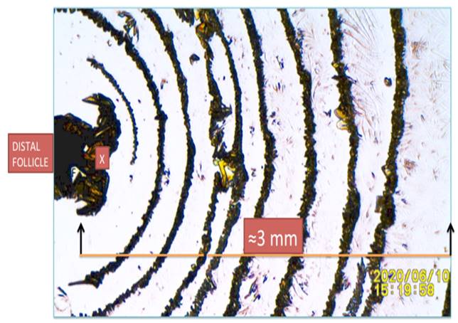
Range of Hair Follicle Electromagnetic Radiation Shown
by Full Absorption of EMRs.
Figure 4
|
Figure 4 Hair follicle magnetic waves range shown as semicircular shapes or Potassium Ferricyanide crystals. X: Crystallization Backguards Suction of hair follicle’s molecules. Note Reproduced
from: Abraham A. Embi Bs. (2018). “THE HUMAN HAIR FOLLICLE PULSATING
BIOMAGNETIC FIELD REACH AS MEASURED BY CRYSTALS ACCRETION.” International
Journal of Research - Granthaalayah, 6(7), 290-299. https://doi.org/10.5281/zenodo.1341349. |
The Fifth- The Shephers Hook Genesis
During the years, in vitro images from experiments of hair follicles on glass slides and covered by different liquids, such as food coloring, calcium carbonate and cyano compunds, all showed truncated magnetic fields unable to fully surround the follicle Figure 5, Figure 6, Figure 7 Again, the inability of the hair magnetic fields from totally surrounding both sides of the hair shaft was displayed. One more time, a mechanism for the truncated signals was unknown Abraham and Embi (2018)
Human Hair Eelectromagnetic Energy Unable to Fully
Circumvent Follicle
Figure 5

|
Figure 5 Freshly plucked scalp human hair covered by calcium carbonate (from my dental plaque) on a glass slide diluted in vinegar after evaporation. Black Arrow: Showing particles unable to penetrate opposite side of follicl |
Follicle on slide covered by drops food coloring Showing
Truncated EMRs
Figure 6

|
Figure 6 Hair follicle in food coloring diluted in water. Showing (Orange Line) Black Arrow: Particles unable to penetrate contralateral side of shaft |
Hair Immersed in Liquid Potassium Ferricyanide Showing
EMRs Unable to Reach Both Sies of Shaft.
Figure 7

|
Figure 7 Hair follicle covered by diluted Potassium Ferricyanide (after evaporation) showin Red Arrow: Semicircular Potassium Ferricyanide crystals (reflecting EMFs) unable to penetrate or continue exiting via a contralateral shaft |
The Sixth- Inference of Hair Shaft’s BipolarityUnilateral Shaft Electromagentic Radiation Triggering Unilateral Blood Coagulation
The bipolar electrical property of the shaft is inferred by using fresh blood on a glass slide. Figure 8 This inference was consciously recognized by this author after reading a publication by DeLangis and Yen (1986) where they state “The majority of the particles within the blood are negatively charged. Although the intima of the vascular system is negatively charged with respect to the adventitia of the vessel, trauma to the vessel will cause the negative charge to become zero or positive with a concomitant thrombosis at that point”. This finding infers that a negative charge would repel and a positive one will attract blood tissue ensuing coagulation.
Example of Bipolar Electrical Charges of Shaft Effect
on Blood
Figure 8

|
Figure 8 Microphotopraph depicts detached hair shaft outline. A=
non-coagulated blood S= Hair shaft
C= Coagulated blood. (-) =
Negative pole RBC repulsion. (+) = Positive pole attracting RBCs
(coagulation). Note Reproduced from
Abraham A. Embi Bs. (2018). “HAIR AND BLOOD ENDOGENOUS LOW LEVEL BIOMAGNETIC
FIELDS CROSS-TALK EFFECTS ON FIBRIN INHIBITION AND ROULEAU FORMATION.”
International Journal of Research - Granthaalayah, 6(11), 200-208. https://doi.org/10.29121/granthaalayah.v6.i11.2018.1118
|
Additional Figure Showing Unilateral
Bioelectromagnetical Radiation of the hair shaft.
Figure 9

|
Figure 9 Hair shaft sanwiched between glass slides and covered by drops potassium Ferrocyanide in solution. Showing shaft unilateral discharge of elecromagnetic energy. Black Arrow: Direction of forces X: Shaft side void of energy |
4. SUMMARY AND DISCUSSION
The introduction of a tabletop optical microscopy method able to detect plant and animal tssue EMRs facilitated the introduction of a mechanism explaining the bipolar property of the hair shaft. This presentation could be summarized as follows:
· The introduction of a tabletop microscopy method facilitated a demostration of the hair follicle’s unique display of electromagnetism not fully surrounding the hair follicle.
· This phenomenon was described as “The Sheperds Hook Effect”
· The initial interpretation stated in the tabletop method stated by the authors (I being one of them) emphasized the attraction of fine iron particles as the main factor for the images, it was then concluded “that the intrinsic metabolic activity contribute an (electromagnetic field) EMF which not only attracts the fine iron particles alowing static as well as streaming images reflecting the electromagnetic fields emanating from plant and anima tissues, respectivelly”
· Only when the Potassium Ferricyanide Anisotropy was realized as having “full absorption of incoming electromagnetic radiation”, is that the absence of crystals in the side of the shaft was correlated with a having a negative charge; and the opposite showing crystals depositions having a positive one, thus identifying the shaft’s bipolarity.
· The unilateral presence of electromagnetic radiation in the hair shaft was obtained in n=6 experiments.
· Figure shown below confirms the above by showing Potassium Ferricyanide unilaterally absorbing electromagnetic radiation (by the presence of one-sided crystals) Figure 10 below.
· Also presented is an electromagnetic image of an in toto tweezers plucked hair follicle. Two novel sequential techniques are needed for this demonstration. The first, the time limited property of a fresh blood smear (approximately 60 seconds). During the “wet stage” the intrinsic blood properties are preserved, i.e.: rejection of foreign materials Abraham and Embi (2019), Embi (2018), Embi (2019)
Hair on Slide Covered by Potassium Ferricyanide.
Hair Physically Removed by Tweezers Showing Absence of Crystals in Negative Side. For details scan QR Code below.
Figure 10

|
Figure 10 The Genesis of The Shepherds Hook Pattern and The Unilateral Biomagnetism of the Human Hair Shaft |
· Plucked scalp hair mounted on a glass slide and covered by drops of Potassium Ferricyanide (KFe3). After evaporation, notice the unilateral presence of biomagnetism of the hair shaft expressed by the triggering KFe3 crystals. Also, the presence of crystals due to biomagnetism surrounding the hair follicle.
· Image reproduced from: Embi (2021). Some curious findings hair follicles bioelectromagnetic radiation expressed as light displacing matter in its path and the contralateral emission of magnetic fields found in the hair shaft. International Journal of Research - GRANTHAALAYAH, 9(7), 334. doi: 10.29121/granthaalayah.v9.i7.2021.4114
· Human Hair Electromagnertic Imprint Showing Gap Resulting in the Shepherd’s Hook Genesis. Seen in Figs
· First Introduced in 2021
EXHIBIT III
Figure 11

|
Figure 11 Exhibit
III. Showing sequential images from video-recordings outlining the human hair
external electromagnetic radiation. Black Arrows: Notice the narrowing shown
between the distal follicle and the bulb also showing a gap in energy
continuity. This gap is theorized to induce the bipolar nature of the shaft
(+-). TIBS= Temporary In Vivo For video detais link to:
https://youtu.be/LLz43yAbpg0 Or scan
QR Code in left side of figure. For
additional details link to: Embi AA (2021) INTRODUCING GAP IN HAIR FOLLICLE
ELECTROMAGNETISM AS MECHANISM FOR THE PRESENCE OF BIPOLAR ELECTRICAL CHARGES
INHERENT IN THE HUMAN HAIR SHAFT DOI: 10.29121/granthaalayah.v9.i9.2021.4260 |
5. CONCLUSION
Of relevance is the demonstration of the hair shaft as a bipolar (+−) extension of the DC currents emitted by the hair follicle.
The freshly plucked in toto human hair is a bioelectromagnetic cohesive unit; the follicle as a DC current generator; and the shaft as its bipolar electrical outlet, thus supporting and duplicating with a table-top microscopy method Scherlag et al. (2016) the seminal work done by Cohen et al. in the early 1980’s Cohen et al. (1980)
CONFLICT OF INTERESTS
None.
ACKNOWLEDGMENTS
None.
REFERENCES
Abraham, A. Embi, B. S. (2018). The Human Hair Follicle Pulsating Biomagnetic Field Reach As Measured By Crystals Accretion. International Journal of Research - Granthaalayah, 6(7), 290-299. https://doi.org/10.29121/granthaalayah.v6.i7.2018.1309
Abraham, A. Embi, B. S. (2018). The Shepherds Hook Phenomenon Pattern of Hair Roots A Demonstration of Comparative Biolectromagnetism Between Human Hairs And Mouse Whiskers By Means of The Photoelectric Effect. International Journal of Research - Granthaalayah, 6(7), 317-326. https://doi.org/10.29121/granthaalayah.v6.i7.2018.1312
Abraham, A. Embi, B. S. (2019). Energy Detection in The Form of Light Radiation At End of Human Blood Coagulation Cascade- The Optical Absorption of Water Vs. Fibrin Burst Energy Release. International Journal of Research - Granthaalayah, 7(9), 200-212. https://doi.org/10.29121/granthaalayah.v7.i9.2019.602
Baranov, D. G. Edgar, J. H. Hoffman, T. Bassim, N. Caldwell, J. D. (2015). Perfect interferenceless absorption at infrared frequencies by a van der Waals crystal. https://doi.org/10.1103/PhysRevB.92.201405
Cohen, D. Palti, Y. Bn, C. & Sj, S. (1980). Magnetic Fields Produced By Steady Currents In The Body. Proc. Natl. Acad. Sci, 77(3), 1447-1451. https://doi.org/10.1073/pnas.77.3.1447
DeLangis, P. A. Yen, T. F. (1986). Electronic antihemocoagulation. Biomater Med Devices Artif Organs, 14(3-4), 195-225. https://doi.org/10.3109/10731198609117543
Embi, A. A. (2018). Biomagnetism as Factor in Red Blood Cells Deformation. International Journal of Research - Granthaalayah, 6(12), 46-57. https://doi.org/10.29121/granthaalayah.v7.i1.2019.1076
Embi, A. A. (2018). Hair and blood endogenous low level biomagnetic fields cross-talk effects on fibrin inhibition and rouleau formation. IJGR, 6(11), 200-208. https://doi.org/10.29121/granthaalayah.v6.i11.2018.1118
Embi, A. A. (2019). Expanding the role of magnetic fields in red blood cells deformations : Demonstration of paramagnetic and diamagnetic fields. IJRG, 7(2), 214-220. https://doi.org/10.29121/granthaalayah.v7.i2.2019.1026
Embi, A. A. Jacobson, J. I. Sahoo, K. Scherlag, B. J. (2015). Demonstration of Inherent Electromagnetic Energy Emanating from Isolated Human Hairs. Journal of Nature and Science, 1(3).
Embí, A. (2016). Demonstration of the Human Hair Shaft as Transmitter/Receiver of Electromagnetic Forces. Journal of Nature and Science, 2, 191.
Figgis, B. N. Gerloch, M. Mason, R. and Nyholmthe, R. S. (1969). The Crystallography and paramagnetic anisotropy of potassium ferricyanide. https://doi.org/10.1098/rspa.1969.0031
Scherlag, B.J. Sahoo, K. Embi, A. A. (2016). A Novel and Simplified Method for Imaging the Electromagnetic Energy in Plant and Animal Tissue. Journal of Nanoscience and Nanoengineering, 2(1), 6-9.
Schneider, M. R. Schmidt-Ullrich, R. Paus, R. (2009). The hair follicle as a dynamic miniorgan. Curr Biol, 19(3), 132-142. https://doi.org/10.1016/j.cub.2008.12.005
 This work is licensed under a: Creative Commons Attribution 4.0 International License
© Granthaalayah 2014-2022. All Rights Reserved.


