
|
|
|
|
NTI-BACTERIAL, CYTOTOXICITY, AND ANTIOXIDANT PROPERTIES OF THE ISOLATED FLAVONOIDS EXTRACT FROM WHITE DRAGON FRUIT (Hylocereus undatus) PEELS AND FLESH
Athena Gabrielle R. Foronda 1![]()
![]() ,
Dr. Elsa L. Cajucom 2
,
Dr. Elsa L. Cajucom 2![]()
![]()
1 Saint
Mary’s University Junior High School, Bayombong, Nueva Vizcaya, Philippines
2 Director,
Center for Natural Sciences, Saint Mary’s University, Bayombong, Nueva Vizcaya,
Philippines
|
|
|
ABSTRACT |
|
|
The presence
of flavonoids in white pitaya’s flesh and peel was confirmed using the Thin
Layer Chromatography Screening method. Flavonoid was extracted using solvent
fractionation and then subjected to the three assays. Using the Kirby-Bauer
method, better antibacterial activity against E. coli was found using 100%
peel extract, partially active at 10.28 mm. The flesh flavonoid extract was
inactive against both E. coli and S. aureus at concentrations of 100%, 75%,
and 50%. The flavonoid peel and flesh have the potential as anticancer agents
since they are active, toxic, and potent, with LC50 of less than 1000 ppm in
Brine Shrimp Lethality Assay. Using a UV VIS spectrophotometer at 517 nm,
both the flesh flavonoid extract and the peel flavonoid extract have
antioxidant potential. However, the flesh with a percent Radical Scavenging
Activity close to the catechin control is a better antioxidant. This study
proved that the peel and flesh of White Pitaya, particularly its flavonoid,
have medical benefits, can be a potential source of income for farmers, and
decrease waste in the environment. |
|||
|
Received 21 February 2023 Accepted 24 March 2023 Published 10 April 2023 Corresponding Author Dr. Elsa
L. Cajucom, ecajucom@smu.edu.ph DOI 10.29121/ijetmr.v10.i4.2023.1305 Funding: This research
received no specific grant from any funding agency in the public, commercial,
or not-for-profit sectors. Copyright: © 2023 The
Author(s). This work is licensed under a Creative Commons
Attribution 4.0 International License. With the
license CC-BY, authors retain the copyright, allowing anyone to download,
reuse, re-print, modify, distribute, and/or copy their contribution. The work
must be properly attributed to its author.
|
|||
|
Keywords: Flavonoids,
White Pitaya, Solvent Fractionation, Kirby-Bauer Method, UV-Vis Spectrophotometer |
|||

1. INTRODUCTION
Flavonoids are a group of organic substances that are known to have good effects on man’s health, this is the reason why research and development are now emerging towards finding its presence in different kinds of fruits which they are known to abound, and their applications to benefit man.
Hylocereus undatus, or white pitaya as it is commonly known, is a type of cactus that is often grown as a decorative plant and also as a fruit crop. It has red skin with green scales, white flesh which is juicy in flavor, and tiny black seeds embedded in it. This fruit is widely planted in hot regions, especially southeast Asian countries, and usually, flowers start from the month of May to August. White pitaya is in season starting end of April but has its peak around June and July until November, but it can extend up to December Gabriel et al. (2015).
Thi-Thuy-Hai Luu et al. (2021) reviewed pitaya’s health benefits and nutrients as well as its sustainable development under climate changes in Vietnam. Both parts of H. undatus and H. p rhizus namely stems, flowers, peels, and pulps contain bioactive compounds such as betalains, flavonoids, polyphenols, terpenoids, steroids, saponins, alkaloids, tannins, and carotenoids. These are proven effective, healthier, safer, and sustainable alternatives to synthetic drugs for the treatment and prevention of many diseases such as diabetes, cancer, obesity, hyperlipidemia, and pathogenic agents such as viruses, bacteria, and fungi.
The secondary metabolites of phenolic nature including flavonoids are responsible for a variety of pharmacological activities Pandey and Kumar (2013). Functional hydroxyl groups in flavonoids mediate their antioxidant effects by scavenging free radicals and/or by chelating metal ions which could be crucial in the prevention of radical generation damaging the target biomolecules since flavonoids have the ability to induce human protective enzyme systems. A number of studies have suggested the protective effects of flavonoids against many infectious (bacterial and viral diseases) and degenerative diseases such as cardiovascular diseases, cancers, and other age-related diseases. Flavonoids are the most common and widely distributed group of plant phenolic compounds, occurring virtually in all plant parts, particularly the photosynthesizing plant cells. Fruits and vegetables having flavonoids have been reported as cancer chemopreventive agents. Flavonoids have long been reported as serving multiple functions in plants. Various abiotic and biotic factors help in the generation of reactive oxygen species (ROS) in plants leading to oxidative stress. Fruits and vegetables are natural sources of flavonoids. The medicinal efficacy of many flavonoids as antibacterial, hepatoprotective, anti-inflammatory, anticancer, and antiviral agents are as well established.
The antibacterial activity of flesh and peel methanol fractions of red pitaya, white pitaya, and papaya on selected food microorganisms was conducted by Abdul Hamid et al. (2012). The methanol extract of the flesh of the fruit showed a broad spectrum of activity against all the Gram-positive bacteria. There was no inhibitory activity on Listeria monocytogenes, Klebsiella, Pseudomonas aeruginosa, and Salmonella entrica typhi of the white pitaya flesh extract. Gram-negative and Gram-positive bacteria are an indication that fruit extracts are a potential source for the production of drugs with a broad spectrum of activity against bacteria (Gram-positive and negative) and can be used in the treatment of infectious diseases caused by resistant microorganisms. Furthermore, Abdul Hamid et al. (2012) also studied the antibacterial property of Hylocereus polyrhizus and Hylocereus undatus peel extracts, using different alcohol extraction.
Hylocereus undatus fruits obtained a higher nutritional value compared to Hylocereus ocamponis fruits. The predominant mineral in both fruit species was potassium. It was revealed in the study that sodium was not found in Hylocereus undatus fruits. Higher antioxidant activity was shown by the pulp of both species. The pulp of the H. ocamponis presented the highest levels of betalains and ascorbic acid revealing higher antioxidant activity which may be linked with the synergistic effect of the different metabolites. The ascorbic acid content determined in their study was lower than the concentration reported by Vaillant et al. (2005) using the three varieties of Hylocereus species cultivated in Nicaragua. This may be accounted for the CAM metabolism in the fruits of Hylocereus which favored the formation of malic acid compared to that of other organic acids. Therefore, results indicated that the pitaya fruits have a higher antioxidant potential than the fruits of some cacti such as the prickly pear (Opuntia ficus-indica) from yellow, red, and white cultivars.
The pulp of H. undatus fruits presented a higher
nutritional value than the pulp of H. ocamponis fruit due to a higher
content of protein, lipids, N, P, K, Ca, Mg, B in the mesocarp, and oleic acid
along with linoleic acid in the seeds. In contrast, the pulp of H. ocamponis
presented the highest nutraceutical potential given the higher betalain content
and antioxidant activity, despite being a fruit of lower commercial demand and
consumption Castillo-González
et al. (2020).
Chemometric analysis indicated phytochemical content-activity relationships when different species and varieties of pitaya fruits were compared in terms of their bioactivity and cytotoxicity Pawel et al. (2012). Based on their findings, the high antioxidant activity of red pitaya was observed. Yellow pitaya showed cytotoxic, but no antioxidant activity. Red pitaya was selectively cytotoxic to gastrointestinal cancer cells.
Ayub Md Som et al. (2018) conducted a study on the extraction of foliage and peels of Hylocereus undatus. Both chloroform and methanol solvent extractions indicated that the peels have hig h phenolic content compared to the foliage. Using chloroform solvent, it was proven that peels contained higher antioxidants than foliage since the percentage of free radical scavenging activity was lower in peels than in foliage. Meanwhile, the antioxidant activity was lower in peels but higher in foliage by using methanol solvents. It was confirmed through the DPPH assay that the antioxidant activity using chloroform extraction was higher compared to methanol extraction.
The presented studies on Hylocereus undatus then revealed its potential anti-oxidant capability and its anti-microbial activity. Several studies also have been made in order to test for the nutritional, cytotoxic, antioxidant, and antibacterial properties of dragon fruit. Thus, this investigation was focused on the isolation of the flavonoid extract and testing its ability to kill microorganisms, particularly S. aureus and E. coli, and its potential as an anti-oxidant, and as a cytotoxic drug. This study sought to find a solution to the increasingly adverse effects that artificial chemicals due on the human body. Using both flesh and peels of the white pitaya could be a good step towards an eco-friendly planet, removing waste and advocating the use would also help the community to have additional livelihood especially now that agritourism pushes towards growing pitaya plants, particularly in Isabela and Nueva Vizcaya. Finally, this research would be a great contribution to the very limited studies on white pitaya flavonoid extracts from peels and flesh. Results of this study shall be added to the long list of literature performed on H. undatus, but specifically more useful in providing information regarding the flavonoid content of H. undatus, peel, and flesh through ethanol solvent fractionation method, and its activity as an antibacterial, antioxidant, and as a cytotoxic agent.
2. MATERIALS AND METHODS
2.1. Materials
The
gathering of White Pitaya was done at Kasibu, Nueva Vizcaya. The fruits
collected were washed thoroughly and placed in the refrigerator for storage,
until ready for use.
2.2. Extraction
The
fruit peel and flesh extract were prepared at the SMUJHS Laboratory The peels
were separated from the flesh. The peels were cut into small pieces and washed
with distilled water. Then it was oven-dried at 60 oC until ready
for use and ground. Ethanol was added and set aside for 48 hours before placing
it in a water bath at 60 oC for 2 days. The flesh was washed with
distilled water, pureed using a blender, and strained using cheesecloth. The
addition of 95% ethanol in a 1:1 dilution was made. After which it was placed
in a water bath until fully extracted.
2.3. Phytochemical Screening
The
detection of the presence of secondary metabolites was determined using Thin
Layer Chromatography. Samples of the plant extract were spotted onto the TLC
silica-gel plates 7 x 4 cm and were developed in the acetate-methanol (7:3)
mixture in the developing chamber. The spots for a certain metabolite were
visualized on the TLC plates and were exposed to UV light and a hot plate to
check the separation of the different compounds. The presence of Phenols,
Sterols, Triterpenes, and Essential Oils was determined through Vanillin-sulfuric
acid reagents. On the other hand, KOH-MeOH was used to test the presence of anthraquinones,
coumarins, and anthrones. Potassium
ferricyanide-ferric chloride reagent was used to detect phenolics compounds and
tannins. Also, Dragendorff’s reagent was
used to spot alkaloids while Antimony (III) chloride was used to detect the
presence of flavonoids. These procedures were conducted based on the method
described by Guevara (2005).
2.4. Flavonoid Extraction
Powdered
dried peels and pureed flesh were soaked in ethanol using the solvent fractionation
method to remove the fats, oils, terpenes, waxes, and the other constituents of
the powdered dried leaves. The ethanol portion was then discarded. To extract
the crude flavonoid of the plant extract, an alcohol mixture consisting of
methanol and ethanol at a 1:1 ratio was used. This was then concentrated in a
water bath at 45-50 oC. The crude extract was separated into a
saturated aqueous tartaric solution and ethyl acetate, in a 1:1 ratio. Neutral
and weakly basic flavonoids were then contained in the ethyl acetate layer. The
ethyl acetate fraction was concentrated. The aqueous layer was then neutralized
with sodium carbonate and extracted again with ethyl acetate. The organic layer
contained the basic flavonoids, while the aqueous layer contained the
quaternary ammonium ions. To determine if there is the presence of secondary
metabolites in the peel and the flesh extracts, the organic layer was subjected
to thin-layer chromatography. The phytochemical screening procedure was done to
characterize the isolated flavonoids. The visualizing agents are FeCl3
and K4 (FeCN)6.
2.5. Antibacterial Assay
Agar
plates were prepared using 28 g of Nutrient Agar (NA), and 1000 mL of distilled
water. The agar mixture was placed in an Erlenmeyer flask and allowed to
dissolve on a hot plate at 420 °C while doing continuous stirring until totally
dissolved. The Petri dishes used were autoclaved at 121 °C. Afterward, the agar
mixture was poured into the sterile petri plates, 15-20 mL of nutrient agar per
plate. Then allowed to solidify for 5 minutes.
The
previously-prepared agar plates were allowed to come to room temperature. If
the visible liquid was present, it was inverted to drain excess liquid from the
agar surface and evaporate. Each plate was labeled with the organism to be
tested, including the concentrations of the extracts, 100%, 75%, 50% positive
control, and negative control.
A
sterile cotton swab was dipped into the previously-prepared inoculum of S. aureus. The swab was streaked three
times over the entire agar surface. The plate was rotated approximately 60
degrees to ensure an even distribution of the inoculum. The swab was discarded.
This was again done for the E. coli.
The lid was left slightly ajar. Then permitted to sit at room temperature for
at least 5-10 minutes, for the surface of the agar plate to dry before
proceeding to the next step.
Extracts
at a concentration of 100%, 75%, and 50%, including the positive control, were
loaded on a 6 mm sterile disk, and allowed to diffuse for 5 minutes. Using the
disks with different concentrations of extracts from peels and flesh, and also
a commercially-prepared antibiotic, the following were done: The inoculated
nutrient agar plate was placed on a flat surface and the lid was removed. Using
forceps, a disk with 100% extract was taken and lightly placed on top of the
agar plate. Disks of the same concentration were strategically placed on the
agar plate. It was again done for the 75%, 50%, positive control (Streptomycin)
and negative control (extracting agent), and both for S. aureus and E. coli.
When the disks were all in place, the lid was replaced, and the plates were
inverted and placed in a 37 °C incubator for 24 hours before reading. After
incubation at 35 °C for 24 hours, the inhibition zone formed around the disk
was measured using a digital Vernier caliper. Results were recorded and
compared.
2.6. Cytotoxicity Test
The
cytotoxicity method was adapted from Olowa and Nuneza (2013), with slight modifications. Cytotoxicity
assay was done by rearing brine shrimps on a salt-water solution to imitate the
seawater environment (3.8 g sodium chloride-100 mL distilled water). The samples
were placed in an improvised two-chambered container. One of the chambers was
kept dark for the eggs and the other chamber served as a catching chamber for
the nauplii. The shrimps were allowed to hatch and mature as nauplii (larva)
for two days. After two days, when the shrimp larvae are ready, 1 mL (1000 ppm,
500 ppm, 250 ppm, 125 ppm, and 62.5 ppm) of the plant extract was added to each
of 15 well plates using 24 well plates and 10 brine shrimps were introduced
into each well. The set-ups for the concentrations were done in triplicate.
Thus, there were a total of 30 shrimps per serial dilution. The well plates
were left uncovered under the light/lamp. The mortality rate of brine shrimps
was recorded every 3-hour interval within 24 hours. To obtain the value for %
mortality by the plant extract, this equation was utilized:
%Mortality = No. of
dead brine shrimps x 100
No. of initial live
brine shrimps
Using
Probit Analysis, the lethality concentration (LC50) was assessed at
95% confidence intervals. LC50 of less than 1000 ppm was considered
as potent (active). The determination of the potency of substances tested for
cytotoxicity assays was based on LC50 values. LC50 value of less
than 1000 ppm are significantly potent or toxic (lethal) while an LC50
value of greater than 1000 ppm is non-toxic Meyer et
al. (1982).
2.7. Antioxidant Assay
The
antioxidant assay was assessed using the DPPH assay used by Kolak, et al, 2006.
To make a stock solution from the concentrated extract, an aliquot was taken to
make 1000 ppm dilution and 1000 ppm of Catechin as control (1 mg/mL). In a
separate plastic cuvette, one mL of prepared stock solution was mixed with 4 mL
of 0.1 nM DPPH solution. Reactions were done in triplicate. The prepared
mixtures were incubated in the dark at 37oC for 30 minutes. Using a
UV-VIS spectrophotometer, the
absorbance readings were monitored at 517 nm. A lower absorbance of the
reaction mixture indicated higher free radical scavenging activity. The ability
to scavenge the DPPH radical was calculated using the formula:
%
Radical Scavenging Effect = [ (Acontrol - Asample) / Acontrol ] x 100
where Acontrol is the DPPH without the test sample or the absorbance of the control while Asample is the absorbance of the test sample containing the mixture of the DPPH and the sample. Catechin was used as the positive control.
3. RESULTS AND DISCUSSION
3.1. Phytochemical Screening
Phytochemical
screening was made on the white pitaya fruit and peel extract, before doing the
solvent fractionization method through the separatory funnel. This was done in
order to identify the secondary metabolites present in each of the plant
extracts. Phytochemical screening was done using thin-layer chromatography
(TLC). The white pitaya peel extract (WPPE) contained essential oils, fatty
acids, sugars, phenols, anthrones, tannins, flavonoids, alkaloids, steroids,
and coumarins; while the WPFE contained essential oils, fatty acids,
triterpenes, sugars, phenols, anthrones, tannins, flavonoids, alkaloids, and
steroids. Results revealed that the secondary metabolites present in both
extracts were essential oils, fatty acids, sugars, phenols, anthrones, tannins,
alkaloids, steroids, and flavonoids. It is notable that triterpene was not
detected in WPPE. For WPFE, coumarin was not screened. The solvent fractionation
method was used to extract the flavonoids as the subject of the study. Table 1
Table 1
|
Table 1 Phytochemical
Screening Results |
|||||||||||
|
Sample Description |
Essential Oils |
Fatty acids |
Sugars |
Phenols |
Anthrones |
Tannins |
Flavonoids |
Alkaloids |
Steroids |
Triterpenes |
Coumarins |
|
White Dragon
Fruit Peel Extract |
+ |
+ |
+ |
+ |
+ |
+ |
+ |
+ |
+ |
- |
+ |
|
White Dragon
Fruit Flesh Extract |
+ |
+ |
+ |
+ |
+ |
+ |
+ |
+ |
+ |
+ |
- |
3.2. Antibacterial Assay
Preparations
of different concentrations of the White Pitaya Peel Flavonoid Extract (WPPFE)
and the White Pitaya Flesh Flavonoid Extract (WPFFE), namely 100%, 75%, and 50%
were made and subjected to Kirby-Bauer Disk Diffusion (KBDD) method to screen
the in vitro antimicrobial activity using nutrient agar plates (NA), together
with the positive control (Streptomycin) and the negative control (ethanol).
Through the SMU Center for Natural Sciences Research Laboratory which conducted
the Antibacterial Assay, the results were as follows: Table 2
Table 2
|
Table 2 Antibacterial Assay Results |
||||||||
|
|
Zones of Inhibition (In mm) |
|||||||
|
Disks |
Gram-positive S. aureus |
Gram-negative E. coli |
||||||
|
|
Trials |
Trials |
||||||
|
Peel extract |
1 |
2 |
3 |
Mean |
1 |
2 |
3 |
Mean |
|
100% |
10.04 mm |
9.99 mm |
9.44 mm |
9.82 mm |
10.56 mm |
10.06 mm |
10.23 mm |
10.28 mm |
|
75% |
9.08 mm |
9.05 mm |
9.16 mm |
9.10 mm |
9.22 mm |
9.40 mm |
9.38 mm |
9.33 mm |
|
50% |
7.93 mm |
8.06 mm |
8.15 mm |
8.05 mm |
8.61 mm |
8.02 mm |
8.37 mm |
8.33 mm |
|
Flesh extract |
1 |
2 |
3 |
Mean |
1 |
2 |
3 |
Mean |
|
100% |
8.10 mm |
8.09 mm |
8.10 mm |
8.09 mm |
8.40 mm |
8.09 mm |
8.12 mm |
8.20 mm |
|
75% |
7.12 mm |
7.45 mm |
7.48 mm |
7.35 mm |
7.56 mm |
7.81 mm |
7.70 mm |
7.69 mm |
|
50% |
6.90 mm |
7.07 mm |
7.15 mm |
7.04 mm |
6.35 mm |
7.23 mm |
7.13 mm |
6.90 mm |
|
Control |
|
|
||||||
|
Positive |
1 |
2 |
3 |
Mean |
1 |
2 |
3 |
Mean |
|
Streptomycin |
|
|
||||||
|
100% |
23.37 mm |
24.39 mm |
23.21 mm |
23.66 mm |
||||
|
75% |
23.37 mm |
24.39 mm |
23.21 mm |
23.66 mm |
||||
|
50% |
23.37 mm |
24.39 mm |
23.21 mm |
23.66 mm |
||||
|
Streptomycin |
|
|
||||||
|
100% |
22.13 mm |
23.76 mm |
23.84 mm |
23.24 mm |
||||
|
75% |
22.13 mm |
23.76 mm |
23.84 mm |
23.24 mm |
||||
|
50% |
22.13 mm |
23.76 mm |
23.84 mm |
23.24 mm |
||||
|
Negative |
1 |
2 |
3 |
Mean |
1 |
2 |
3 |
Mean |
|
Extracting agent |
6 mm |
6 mm |
6 mm |
6 mm |
6 mm |
6 mm |
6 mm |
6 mm |
|
Legend: <10mm=inactive; 10-13 mm=partially active;
14-19 mm=active; >19mm=very active |
||||||||
For
the antibacterial assay conducted, it is the WPPFE that exhibits significant
inhibitory effects as compared to the WPFFE. Results show that only the 100%
WPPFE is partially active against E. coli with a mean zone of inhibition of
10.28 mm but is inactive against S. aureus with a mean zone of inhibition of
9.82 mm, 9.10 mm, and 8.05 mm, at a concentration of 100%, 75%, and 50%,
respectively. WPFFE, at all concentrations, is inactive against both E. coli and S. aureus, with a mean zone of inhibition of less than 10 mm for
the three concentrations. However, all the concentrations of the WPPFE and the
WPFFE exhibit higher zones of inhibition than the extracting agent (ethanol),
the negative control, which is 6 mm in diameter. Thus, there is still the potential
for WPPFE and WPFFE to be used as antimicrobial agent, even if it does not
exhibit the same effectiveness as the commercially prepared antibiotic that was
used as the positive control. In this case, Streptomycin was used instead of
the drug of choice for both E. coli
and S. aureus which are
Trimethoprim/Sulfutamethoxazole and Cloxacillin, respectively.
Based
on the antibacterial assay performed, it revealed that the White Pitaya Peel
Flavonoid Extract (WPPFE) is partially active against E. coli at 100% concentration, but is inactive against S. aureus at 100%, 75%, and 50%. The
White Pitaya Flesh Flavonoid Extract (WPFFE) is inactive against both E. coli and S. aureus at all concentrations. In a study done by Nassar, Hazzah,
and Bakr, in the evaluation of antibiotic susceptibility test results,
published in the Journal of the Egyptian Public Health Association in 2019,
“Muller-Hinton agar is a loose agar that allows for better diffusion of the
antibiotics than most other media. The high deviation of AST results observed
between MHA and NA raises doubts about the reliability of the NA for
susceptibility testing and should not be used as a substitute for MHA.”,
because nutrient agar is a general-purpose medium rather than a standard
susceptibility testing medium. According to Donkor et al. which compared
nutrient agar (NA) and MHA in antimicrobial susceptibility testing of S. typhi and S. aureus isolates, there is an overall discrepancy in
susceptibility results between NA and MHA at 8.9%. Thus, according to the
research, the use of nutrient agar in the Kirby-Bauer method is discouraged due
to the considerable error margin that the nutrient agar may introduce into
susceptibility results.
Based
on the antibacterial testing, the White Pitaya (Hylocereus undatus) Peel
Flavonoid Extract has antibacterial potential for Escherichia coli. It
is partially active in E. coli at 100% concentration but is inactive against Staphylococcus
aureus in all concentrations. The White Pitaya Flesh Flavonoid Extract is
inactive against both E. coli and S. aureus.
3.3. Cytotoxicity Test
The
percent mortality rate for the WPPFE is 100% starting at 3 to 24 hours at 1000
ppm, but for 500 ppm, it is 10% from 15 hours to 24 hours. The percent
mortality rate was computed to ensure the death of nauplii is due to the
bioactive compounds present in the flavonoid plant extract. The LC50 of the
WPPFE from 3 to 24 hours are all less than 1000 ug/ml, thus, the plant extract
is potent and toxic, as compared to the WPFFE which was only potent from 6 to
24 hours, while the 500 ppm is only potent and toxic from 21 to 24 hours. Based
on the results and interpretations according to Meyer et al., the LC50¬ of less
than 1000 ppm is toxic (active), while an LC50 of more than 1000 ppm is
non-toxic (inactive), both the WPPFE and the WPFFE have cytotoxic properties
due to the bioactive compound present, flavonoid, indicating the potential to
be an anticancer agent. Table 3, Table 4
Table 3
|
Table 3 Cytotoxicity Assay (Peel) Results |
||||||||
|
Percent Mortality Rate (%) (Peel) |
||||||||
|
Concentration |
3hrs |
6hrs |
9hrs |
12hrs |
15hrs |
18hrs |
21hrs |
24hrs |
|
1000 ppm |
100 |
100 |
100 |
100 |
100 |
100 |
100 |
100 |
|
500 ppm |
0 |
0 |
0 |
0 |
10 |
10 |
10 |
10 |
|
250 ppm |
0 |
0 |
0 |
0 |
0 |
0 |
0 |
0 |
|
125 ppm |
0 |
0 |
0 |
0 |
0 |
0 |
0 |
0 |
|
62.5 ppm |
0 |
0 |
0 |
0 |
0 |
0 |
0 |
0 |
|
LC50 |
672.04 |
672.04 |
672.04 |
672.04 |
648.49 |
648.49 |
648.49 |
648.49 |
Table 4
|
Table 4 Cytotoxicity Assay (Flesh) Results |
||||||||
|
Percent Mortality Rate (%) (Flesh) |
||||||||
|
Concentration |
3hrs |
6hrs |
9hrs |
12hrs |
15hrs |
18hrs |
21hrs |
24hrs |
|
1000 ppm |
0 |
100 |
100 |
100 |
100 |
100 |
100 |
100 |
|
500 ppm |
0 |
0 |
0 |
0 |
0 |
0 |
10 |
10 |
|
250 ppm |
0 |
0 |
0 |
0 |
0 |
0 |
0 |
0 |
|
125 ppm |
0 |
0 |
0 |
0 |
0 |
0 |
0 |
0 |
|
62.5 ppm |
0 |
0 |
0 |
0 |
0 |
0 |
0 |
0 |
|
LC50 |
Non-toxic |
672.04 |
672.04 |
672.04 |
672.04 |
672.04 |
648.49 |
648.49 |
Based
on the cytotoxicity assay result using the Brine Shrimp Lethality Assay, both
the flavonoid extract of the White Pitaya peel and flesh is toxic. Thus, potent,
and active to kill cancer cells.
3.4. Antioxidant Assay
The
Antioxidant Assay of the WPPFE and the WPFFE using the DPPH
(2,2-diphenyl-1-picrylhydrazyl) method revealed the following table of results:
Table 5
Table 5
|
Table 5 Antioxidant Assay Results |
|||||
|
Sample Description |
Absorbance Reading |
Mean Absorbance |
% Radical Scavenging Activity (RSA) |
||
|
|
1 |
2 |
3 |
|
|
|
Peel Extract |
0.049 |
0.050 |
0.053 |
0.051 |
32.89% |
|
Flesh Extract |
0.031 |
0.030 |
0.028 |
0.030 |
60.53% |
|
Catechin (control) |
0.031 |
0.029 |
0.025 |
0.028 |
63.16% |
Absorbance
reading was made at 517 nm using a UV VIS spectrophotometer. The White Pitaya
Flesh Flavonoid Extract (WPFFE) has a lower absorbance mean of 0.030, as
compared to the White Pitaya Peel Flavonoid Extract (WPPFE) with a mean of
0.051. The mean absorbance of the control (catechin) is 0.028, which is close
to that of the WPFFE at 0.030. Since a lower absorbance translates to a higher
percent Radical Scavenging Activity (RSA), this indicates that the WPFFE has a
higher RSA than the WPPFE. Although there is a difference between their RSA,
both the WPPFE and the WPFFE have antioxidant properties, only, it is higher in
the WPFFE. Thus, both the WPPFE and the WPFFE have the potential to be used as
an antioxidant agents.
Both
the WPPFE and the WPFFE have antioxidant properties, but the WPFFE has a
greater radical scavenging activity (RSA). The 60.53% RSA is close to the
63.16% of catechin (control). Thus, the flavonoid extract for both peel and
flesh have the potential as an antioxidant agent. In a study done in 2020 by
Hernandez-Ramos et al., where the fruits of the H. undatus and the H.
ocamponis were compared as to their nutritional components and
antioxidants, it revealed that there were no differences in the antioxidant
activity in the tissue between both species. According to the study done by Som
et al., when the peels and foliage were compared as to their antioxidant
activity and phenolic content, the peels contained more antioxidants than the
foliage in chloroform solvent extract. The result also showed that in
comparison between the chloroform and methanol solvents used, the methanol
extract showed a high RSA for both foliage and peels as compared to the
chloroform. Thus, the solvent used has an effect on the result of the
antioxidant activity of plant extracts.
Based
on the antioxidant assay, the White Pitaya Flavonoid Extract from the peel and
from the flesh is effective as an antioxidant agent to inhibit the formation of
free radicals, but the flesh has a greater radical scavenging activity.
4. CONCLUSIONS ANd RECOMMENDATIONS
At these times when the environment is in the midst of destruction caused by the numerous chemicals being used, it is just timely that this scientific study is done. It not only answers the antioxidant, cytotoxic, and antibacterial benefits that the White Pitaya gives to man, but it also answers environmental problems such as the accumulation of waste. The peels that are normally thrown away after getting the flesh for food are now revealed to have the potential medicinal properties of the flavonoid it contains.
From the study conducted, it can be concluded that the White Pitaya Flavonoid Extract from the peel, has an antibacterial, cytotoxic, and antioxidant potential, while the White Pitaya Flavonoid Extract from the flesh has cytotoxic and antioxidant potential.
However, future studies are recommended since there is still a potential for the flesh and the peel to be sensitive to other microorganisms. Also, it is recommended that further studies be done on the anticancer potential of WPPFE and WPFFE, since the brine shrimp lethality assay is only a method for screening and fractionation of new bioactive natural products from plants, according to Osamudiamen et al. (2020). Moreover, the extraction of H. undatus flavonoid extract will be done using different solvents, like chloroform and methanol since the solvent used for extraction has an effect on the antioxidant activity.
CONFLICT OF INTERESTS
None.
ACKNOWLEDGMENTS
The researchers would like to give special thanks to all of the people who have helped throughout the whole process of this research project. To the SMU CNS Director, Dr. Cesar T. Medula Jr., and the laboratory assistants of Saint Mary's University; Mr. Regidor Almendral, Mr. Hanson Villanueva, & Mrs. Jolina Mayawin for their incomparable dedication and support throughout the experimentation process. To Dr. Samuel R. Soliven, Director 3 of the Department of Education, for the unparalleled encouragement to the researchers. To Dr. Michelle Rodrigo-Maniquis, Head of the Pathology Department Region 2 Trauma and Medical Center, for the assistance given to the researcher. To Mrs. Claire Dontogan, RMT, Section Head of the Region 2 Trauma and Medical Center Laboratory, for extending help in providing the microbes needed for the assay. To Drs. Marciano and Rachelle Foronda, for the never-ending assistance and support in all aspects that this project needed to succeed.
REFERENCES
Abdul Hamid, A., Mohamad Ghazali, F., Nurmahani, M. M., Osman, A., and Pak Dek, M. S. (2012). Antibacterial Property of Hylocereus Polyrhizus and Hylocereus Undatus Peel Extracts.
Ahmat, N., Azizuddin, N. M., Hamid, H. A. A., and Som, A. M. (2019). A Comparative Study on Foliage and Peels of Hylocereus Undatus (White Dragon Fruit) Regarding their Antioxidant Activity and Phenolic Content. Heliyon 5(2). https://doi.org/10.1016/j.heliyon.2019.e01244.
Ali, A. M., Bakar, A. A., and Rohin, M. A. K. (2012). Antibacterial Activity of Flesh and Peel Methanol Fractions of Red Pitaya, White Pitaya and Papaya on Selected Food Microorganisms. International Journal of Pharmacy and Pharmaceutical Sciences, 4, 185-190.
Aslanturk, O. (2017). In Vitro Cytotoxicity and Cell Viability Assays: Principles, Advantages, and Disadvantages. A Predictable Risk to Our Actual World. https://doi.org/10.5772/intechopen.71923.
Benson, H. J. (2002). Microbiological Applications : Laboratory Manual in General Microbiology. McGraw-Hill, Boston, MA.
Botha, F. S., Elisha, I. L., McGaw, L. J. et al. (2017). The Antibacterial Activity of Extracts of Nine Plant Species with Good Activity Against Escherichia Coli Against Five Other Bacteria and Cytotoxicity of Extracts. BMC Complement Altern Med 17, 133. https://doi.org/10.1186/s12906-017-1645-z.
Bronze, M. S., and Madappa, T. (2019). Escherichia Coli (E Coli) Infections.
Cai, Y., Liu, T., Luo, H., and Peng, Z. (2014). Chemical Composition and in Vitro Evaluation of the Cytotoxic and Antioxidant Activities of Supercritical Carbon Dioxide Extracts of Pitaya (Dragon Fruit) Peel. Chemistry Central Journal 8(1), 1. https://doi.org/10.1186/1752-153X-8-1.
Castillo-González, A. M., García-Mateos, M. del R., Hernández-Ramos, L., Nieto-Ángel, R., and Ybarra-Moncada, C. (2020). Fruits of the Pitahaya Hylocereus Undatus and H. Ocamponis : Nutritional Components and Antioxidants. Journal of Applied Botany and Food Quality 93, 197-203. https://doi.org/10.5073/JABFQ.2020.093.024.
Chandra, S. R., Diwan, A. D., and Panche, A. N. (2016). Flavonoids : An Overview. https://dx.doi.org/10.1017%2Fjns.2016.41.
Cheng, A., Guerrant, R. L., Pitout, J., and Thielman, N. M. (n.d.). Escherichia Coli.
Gabriel, M. L. S., Pascua, L. T., and Pascua, M. E. (2015). Dragon Fruit Production and Marketing in the Philippines : Its Status, Constraints and Prospects.
Gnanamani, A., Hariharan, P., and Paul-Satuaseela, M. (2017). Staphylococcus Aureus : Overview of Bacteriology, Clinical Diseases, Epidemiology, Antibiotic Resistance and Therapeutic Approach. Frontiers in Staphylococcus Aureus. https://doi.org/10.5772/67338.
Hudzicki, J. (2009). Kirby-Bauer Disk Diffusion Susceptibility Test Protocol.
Janmeda, P., and Veena, S. (2012). Extraction, Isolation and Identification of Flavonoid from Euphorbia Neriifolia Leaves. Arabian Journal of Chemistry, 10(4), 509-514.
Kolak, U., Osturk, M., Ozgokce, F., and Ulubelen, A. (2006). Norditerpence Alkaloids from Delphenium Linearilobum and Antioxidant Activity. Phytochemistry 67, 2170-2175. https://doi.org/10.1016/j.phytochem.2006.06.006.
Nassar, M. S. M., Hazzah, W. A., and Bakr, W. M. K. (2019). Evaluation of Antibiotic Susceptibility Test Results : How Guilty a Laboratory Could be? J. Egypt. Public. Health. Assoc. 94, 4 (2019). https://doi.org/10.1186/s42506-018-0006-1.
Olowa, L. F., and Nuneza, O. M. (2013). Brine Shrimp Lethality Assay of the Ethanolic Extracts of Three Selected Species of Medicinal Plants from Iligan City, Philippines. Vol. 2(11), 74-77.
Osamudiamen, P. M, Aiyelaagbe, O. O., Vaid, S., Sangwan, P. L., Ogbesejana A. B., and Saxen, A. K. (2020). Comparative In-Vitro Anticancer and Brine Shrimp Cytotoxic Activities of Mezoneuron Benthamianum Baill. Journal of Medicinal Plants for Economic Development, 4(1). https://doi.org/10.4102/jomped.v4i1.73.
Pandey, A. K., and Kumar, S. (2013). Chemistry and Biological Activities of Flavonoids : An Overview. The Scientific World Journal. https://doi.org/10.1155/2013/162750.
Praciak, A., and Rojas-Sandoval, J. (2015). Hylocereus Undatus (Dragon fruit). Invasive Species Compendium.
Sindhu, K. M. V. (2014). Phytochemical Screening of Plants.
Appendix
Appendix 1
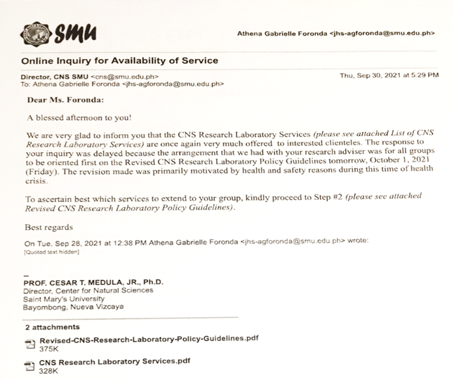
|
Appendix 1 Approval of
the CNS Director to Proceed to Step 2 |
|
|
 This work is licensed under a: Creative Commons Attribution 4.0 International License
© IJETMR 2014-2023. All Rights Reserved.