ShodhKosh: Journal of Visual and Performing ArtsISSN (Online): 2582-7472
|
|
Visual Analysis and Machine Learning–Based Detection of Cardiac Abnormalities from ECG Signals
Priyanka Rane 1![]() , F. Rahman 2
, F. Rahman 2![]() , Rais
A. Mulla 3
, Rais
A. Mulla 3![]()
1 Department
of Computer Engineering Science, Kalinga University, Raipur, India
2 Department
of Computer Engineering Science, Kalinga University, Raipur India
3 Department of Computer Engineering,
Vasantdada Patil Pratishthan’s College of Engineering, Mumbai, India
|
|
|
ABSTRACT |
|
|
CVDs has been
identified as one of the leading causes of mortality in the world and it is
important to make sure that cardiac abnormalities are treated at an early age
to enhance the survival of patient. Electrocardiography represents a
non-invasive test that is used to measure the activity of the heart and
determine abnormal heart rhythm. Hand reading of the ECG signals is however
time consuming and highly skilled in skills especially where a significant
amount of physiological information is required to be read. As of relatively
recent advances in machine learning and biomedical signal processing,
nowadays, automated diagnostic systems capable of detecting cardiac
abnormalities with pretty high degree of accuracy can be developed. This
article study suggests a single-balanced framework of detecting abnormal
patterns of heart beats by visual inspection and machine learning techniques
on ECG signals. The proposed solution includes signal preprocessing methods
in order to remove noise and a line-drift, followed by features extraction
methods that reflect the time-domain characteristics and frequency-domain
characteristics of ECG signals. The visual analytics software will be
utilized to represent the pattern of waveforms and identify the
characteristics of the dataset to understand the variations of the signals
better. They used several classification models including Support Vector
Machines, Rand Forest classifiers, Convolutional Neural Networks, as well as,
Long Short-Term Memory networks to evaluate the applicability of machine
learning models in detection of abnormalities in ECG signals. According to
the results of the experiments, the performance of the deep learning models
is superior to the classical classification algorithms and achieves the
accuracy of more than ninety-seven percent. The visualization methods are
also helpful in the knowledge regarding the model behavior through the
support of demonstrating the performance of classifications and
characteristics of data. The framework developed depicts the effectiveness of
visual-level analysis and machine learning of automated ECG signal
interpretation. Such systems can assist clinicians in detecting cardiac
abnormalities with less effort and allow developing intelligent healthcare
monitoring systems that can potentially carry out cardiac diagnostics in real
time. |
|||
|
Received 20 November 2025 Accepted 13 January 2026 Published 28 March 2026 Corresponding Author Priyanka
Rane, priyanka.rane9333@gmail.com DOI 10.29121/shodhkosh.v7.i2s.2026.7322 Funding: This research
received no specific grant from any funding agency in the public, commercial,
or not-for-profit sectors. Copyright: © 2026 The
Author(s). This work is licensed under a Creative Commons
Attribution 4.0 International License. With the
license CC-BY, authors retain the copyright, allowing anyone to download,
reuse, re-print, modify, distribute, and/or copy their contribution. The work
must be properly attributed to its author.
|
|||
|
Keywords: ECG Signal Analysis, Cardiac Abnormality Detection,
Machine Learning, Biomedical Signal Processing, Arrhythmia Classification,
Deep Learning, Visual Analytics, Healthcare Diagnostics |
|||


1. INTRODUCTION
Cardiovascular diseases are a global health issue and claim thousands of millions of lives annually in both the developing and the developed countries. The timely diagnosis and the correct identification of cardiac abnormalities play a key role in ensuring the decrease of mortality rate and patient outcomes. Electrocardiography is widely used as a procedure in clinical practice because it is a non-invasive technique of monitoring the electrical activity of the heart. ECG signals prove useful in revealing a mandatory information on the heart rhythm, circuits of conduction and condition of the cardiac tissues Jandera et al. (2025). Despite its importance, it can be tedious and time consuming due to the morphological variation of the waveforms and noise or artifact of the signal to be interpreted manually in ECG recordings. These are some of the elements of the waveforms that medical practitioners put into consideration, such as P wave, QRS complex, and T wave of the existence of abnormal cardiac conditions analysis. The problem of uncertainty of variability of patients also contributes to the difficulty of proper interpretation Ayano et al. (2023), as well as variation in recording conditions.
The latest advancements in the area of computational intelligence have allowed one to create the automated ECG analysis systems that can be used to assist clinicians in detecting cardiac abnormalities effectively. Machine learning has been found to be very useful in identifying patterns, classifying and predicting models and hence would be useful in the analysis of biomedical signals. These algorithms are capable of obtaining complex relationship in large amounts of data and locating small shifts in signals which may be challenging to locate in a otherwise manually analyzed process. At the same time, the visual analytics techniques can be applied to provide the intuitive visuals of the patterns of the signals that can guide the researchers and clinicians to understand how the deviations can be visible in the ECG waveforms Basco et al. (2025).
The combination of machine learning with visual signal analysis offers a powerful automated system of cardiac diagnostics. Having high accuracy, such systems may be utilized in dealing with a lot of ECG data and derive meaningful information and discriminate between abnormal heart conditions. Another method of enhancing interpretability to allow clinicians to trace the existence of abnormal patterns and verify prediction of algorithms is visual analysis. The overall healthcare transformation is also contributed to by the development of intelligent ECG diagnostic frameworks that are developed based on data-driven technologies Attia et al. (2021). A series of automated analysis tools has a potential of supporting remote monitoring systems, wearable health devices and telemedicine platforms and enable the continuous monitoring of the cardiac conditions outside the traditional clinical environment. These technologies increase the accessibility to cardiac care and reduce the diagnostic load of health workers. Machine learning-based ECG analysis research is therefore a significant step to a more efficient and scalable healthcare system.
2. Background and Related Work
Changes in these components of the waveforms may be a sign of various cardiac abnormalities like arrhythmias, myocardial infarction, or conduction disorders. In conventional versions of ECG analysis, rule-based algorithms and signal processing techniques are mostly used to identify abnormalities in the recorded signals Chong et al. (2025). The initial automated ECG analysis systems were devoted to the identification of the R-peaks and quantification of the time between the consecutive heartbeats. Algorithms based on signal filtering and peak detection were common to detect the parts of a heartbeat and determine heart rate variability and QRS duration among other parameters. Although these methods yielded valuable information, they were generally restricted by the sensitivity of noise and the variability of the morphology of the waveform in different patients Denysyuk et al. (2023). The performance of automated ECG classification systems has been enhanced by machine learning methods to a great extent. The Support Vector Machines, k-Nearest Neighbor classifier and the random forest models are just some of the algorithms that have been extensively used in the classification of ECG signal. The techniques utilize features obtained off the ECG signals in order to train patterns linked to normal and abnormal cardiac conditions. The commonly used features extraction methods involve the time-domain statistics, frequency-domain features, and the morphological features of the ECG waveforms Goettling et al. (2024).
The ECG signal analysis has also been developed through deep learning techniques. Convolutional Neural Networks have also shown high potential in automatically learning emergent hierarchical features directly out of raw ECGs. Recurrent neural networks and Long Short-Term Memory models have been also used to represent temporal correlations in sequential cardiac signals Pande et al. (2024). With these models, arrhythmias and other heart diseases can be detected with high levels of precision automatically. The recent research has also been conducted into visual analytics in ECG signal interpretation. Visual representation (spectrograms, heatmaps, and waveform plots) helps the researcher to visualize the alterations in the signal and abnormal trends better. The visual examination of the heart optical properties and machine learning algorithms have a positive effect on the readability of the process and the improved understanding of how the classification algorithms are applied to identify an abnormal heart activity He et al. (2025). Amidst many automated ECG classification systems, challenges remain to be obtained in order to attain reliable results when using a large number of patients and recording conditions. These issues need to be solved on the basis of the unified systems, including powerful signal preprocessing, informative feature extraction and advanced machine learning organization. The study of these hybrid approaches remains a major research issue in biomedical signal processing.
3. ECG Signal Acquisition and Dataset Description
The quality of ECG datasets needed to provide the reliable analysis of cardiac abnormalities is the dataset representing physiological cardiac activity. The ECG signals are normally recorded by the electrodes placed on the body surface to measure electrical potentials produced when the heart muscles contract. Normal clinical ECG recordings include many leads, each of which is a measure of electrical activity in another spatial position of the heart. These signals give detailed information with respect to cardiac rhythm and conduction patterns Darmawahyuni et al. (2024). Publicly accessible datasets are typical in machine learning-based ECG analysis research models to train and assess the classification models. The MIT-BIH Arrhythmia Database is one of the most popular datasets that includes annotated ECG records of several patients that had different heart diseases. The data consists of the recordings that were sampled at a rate of 360 Hz and also have the annotations of various types of arrhythmias and abnormal heartbeats Rahman et al. (2023). Such annotations enable researchers to come up with supervised learning models that identify certain cardiac abnormalities.
Table 1
|
Table 1 ECG Dataset Description and Statistics |
||||
|
Dataset |
No. of Records |
Sampling Frequency
(Hz) |
No. of Patients |
Signal Duration
(hours) |
|
MIT-BIH Arrhythmia
Database |
48 |
360 |
47 |
24 |
|
PTB Diagnostic ECG
Database |
549 |
1000 |
290 |
15 |
|
European ST-T Database |
90 |
250 |
79 |
22 |
|
INCART Arrhythmia
Database |
75 |
257 |
32 |
18 |
|
PhysioNet Challenge
Dataset |
8528 |
300 |
400+ |
30 |
The ECG recordings are preprocessed with noise and artifacts which can distort the signal interpretation removed before analysis. The classical sources of noise are interference with muscle activity, electrode motion artifact and power line interference. The signal filtering methods (bandpass filtering and wavelet-based denoising) are frequently used to enhance the signal quality Feyisa et al. (2022).
Another task that constitutes dataset preparation is the balancing of the train sample by different categories of cardiac conditions. Class imbalance is a typical issue with ECG datasets since abnormal heartbeats are not common as compared to normal heartbeats. This problem can be solved by data augmentation and resampling techniques and provide better performance to classification models.Descriptive statistics of the dataset characteristics can answer such questions as how the variability of the signals is and how the cardiac conditions are distributed in the dataset Liu et al. (2021). Mean heart rate, QRS duration, variability of the RR interval among recorded signals are parameters that may allow the researcher to comprehend physiological differences among recorded signals. All these properties also determine the choice of the right feature extraction methods to use in machine learning models.
4. ECG Signal Preprocessing and Feature Extraction
Preprocessing of the ECG signal is a critical consideration in increasing the reliability of automated abnormality detection systems of the heart. Raw ECG signals can be subject to numerous noise and signal distortions and this can negatively affect the quality of the ensuing analysis steps. It can be then concluded that there should be good preprocessing techniques that enhance the quality of signals and ensure that physiological information is stored in an analytical stage Śmigiel et al. (2021).
Filtering of noise can be considered the preprocessing process to a large extent. ECG signals may introduce baseline wander (respiration and electrode movement), high frequency noise (muscle activity and external electrostatic interferences). Digital filtering filters such as bandpass filters are highly applied with the aim of being in a position to filter out such unwanted elements Varagani (2026). The other use of the wavelet-based filtering techniques has been trendy due to their ability to preserve morphology of the signal without noise.
Table 2
|
Table 2 ECG Signal Preprocessing Parameters |
|||
|
Preprocessing Stage |
Method Used |
Parameter Value |
Purpose |
|
Noise Filtering |
Bandpass Filter |
0.5–40 Hz |
Remove baseline wander
and high-frequency noise |
|
Powerline Noise
Removal |
Notch Filter |
50 Hz |
Remove electrical
interference |
|
Signal Normalization |
Min-Max Scaling |
0–1 range |
Standardize amplitude
values |
|
R-Peak Detection |
Pan-Tompkins Algorithm |
Threshold = 0.6 |
Identify heartbeat
cycles |
|
Signal Segmentation |
Windowing |
200 samples |
Extract individual
heartbeats |
After noise removal, precise R-peaks detection in the QRS complex is done. QRS complex is the most salient part of the ECG recording and is used to divide the heartbeat sequences. Many algorithms including the Pan-Tompkins have been commonly used to determine the R-peak position in the ECG signal. After the R-peaks have been identified, the signal is divided into separate cardiac cycles to do additional analysis. The feature extraction process transforms the segmented ECG signals into numbers which can be used by machine learning algorithms. The time-domain values include values of the RR intervals, QS duration, and statistical values such as mean amplitude and variance. Signal transformation methods (i.e. the Fourier transform or the wavelet transform) are used to generate frequency-domain features which state the frequency content of the cardiac activity. The form of the waveforms as described by morphological characteristics are also important indicators of cardiac malfunctions. These features document the attributes of the P wave, QRS intricacy, and T wave which may vary in case of various types of arrhythmia or heart illness. Machine learning models can be employed to give a comprehensive account of ECG signals by employing a number of feature types. Effective feature extraction enhances the discriminative power of classification algorithms, as well as, elevates quantity of detection. The automated diagnostic systems will be more efficient in differentiating the normal and abnormal cardiac conditions because they convert raw signal into information-bearing representations of the signals.
5. Analysis Framework for ECG Patterns
Visual analysis methods are useful in getting good ideas regarding the complicated patterns available in ECG signals. ECG waveforms are presented in graphical form which helps researchers and clinicians to note morphological changes related to cardiac malformations. These visualizations can be used as supplements to machine learning models since it can provide interpretable perspectives of signal properties that might otherwise be unseen using numerical analysis.
Figure
1
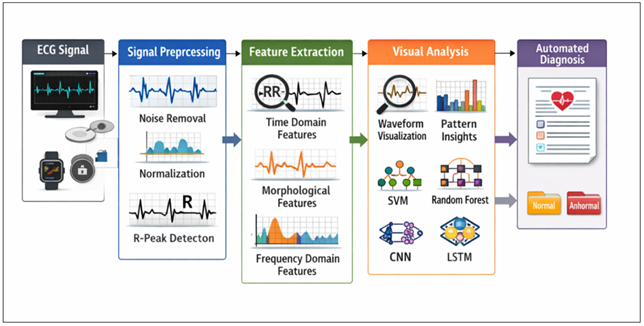
Figure 1 ECG Signal Processing and Machine Learning-Based Automated Cardiac Diagnosis Framework
Figure 1 shows the workflow of automated ECG analysis. Raw ECG signals undergo preprocessing including noise removal, normalization, and R-peak detection. Extracted time, morphological, and frequency features are analyzed using machine learning models (SVM, Random Forest, CNN, LSTM) to enable visual interpretation and automated classification of normal or abnormal cardiac conditions.
Waveform visualization can be defined as the most basic type of ECG signal analysis. The time dependency of ECG signals can be plotted enabling the clinicians to review the arrangement of P waves, QRS complexes, and T waves in the separate cardiac cycles. Such abnormalities may be irregular heartbeat intervals, abnormal QRS durations or inverted T waves, which can be often identified by attentively observing the pattern of the waveforms. Time-frequency representations deliver more information about the ECG signal behavior. Spectrograms represent the variation of frequency content of a signal with time and show patterns of abnormal cardiac activity. Such visualizations come in handy especially in detecting minor signal disparities that might arise at times of arrhythmia. The wavelet transform representations also provide localized signal representations in detail allowing one to determine transient events in the ECG recordings.
Visual analytics models ensure the implementation of numerous visualization tools to assist in the overall interpretation of signals. As an illustration, the visualization of ECG recordings may be analyzed using heatmaps, to indicate high signal variability areas, whereas clustering may be applied to ECG recordings, to indicate any similarity between a specific pattern of heartbeat and a different one. These methodologies will help the scientists in learning how various heart conditions appear in ECGs. Visualization methods can be used to prove the predictions of the model by demonstrating the patterns of signals related to identified abnormalities.
Creation of sophisticated visualization systems is still significant to assist in enhancing automated ECG analysis systems. Visual analytics can be used alongside intelligent healthcare technologies because it can improve their performance through increasing their interpretability and offering an intuitive display of complex biomedical signals.
6. Machine Learning Model for Cardiac Abnormality Detection
It has been established that machine learning algorithms can be very useful in providing automated cardiac abnormality detection using ECG signals. These algorithms can adapt to learning intricate associations among signal features extracted and certain cardiac states. With annotated ECG datasets that have been trained on classification models, automated diagnostic systems are able to detect abnormal pattern related to an arrhythmia and other heart diseases.
Figure
2

Figure 2 Machine Learning Model for Cardiac Abnormality Detection
ECG classification has been a popular subject of use of the traditional machine learning algorithms. SVMs are specifically efficient in dealing with high dimensional feature spaces and may construct optimal decision boundaries among normal and abnormal signal classes.
Table 3
|
Table 3 Extracted ECG Features for Machine Learning Model |
|||
|
Feature Category |
Feature Name |
Description |
Units |
|
Time Domain |
RR Interval |
Time between
successive R-peaks |
ms |
|
Time Domain |
QRS Duration |
Width of QRS complex |
ms |
|
Morphological |
P-Wave Amplitude |
Maximum amplitude of P
wave |
mV |
|
Morphological |
T-Wave Amplitude |
Maximum amplitude of T
wave |
mV |
|
Frequency Domain |
Dominant Frequency |
Peak frequency
component |
Hz |
|
Statistical |
Mean Signal Amplitude |
Average ECG amplitude |
mV |
|
Statistical |
Signal Variance |
Variability in ECG
amplitude |
mV² |
The current developments in the field of deep learning have also enhanced the ECG signal analysis capabilities. Convolutional Neural Networks have the ability to learn hierarchical features representations using raw ECG signals without any human intervention. These models do not need that the features be extracted manually they locate the appropriate signal patterns through convolutional filters. The use of CNN models has been very effective in arrhythmia and other related heart defects detection.
Recurrent neural networks and Long Short-Term Memory also happens to be quite appropriate in the analysis of ECG signals as it allows capturing a temporal dependence of the sequential heartbeat signals. They are time-series models that discover patterns that are found in many cardiac cycles to categorize a more sophisticated condition of arrhythmias into the correct category.
Training of a machine learning model involves optimization of the model parameters using the aid of the labeled ECG datasets. Such techniques as hyperparameter tuning, and cross-validation are used to improve generalization performance. Evaluation metrics include accuracy, precision, recall and F1-score which are quantitative measures of the effectiveness of the model.
The multiple machine learning models can be incorporated further to improve classification. Such methods are the ensemble learning methods which entail the unification of the prophesying of different algorithms to achieve a high degree of steadiness and precision. These mixed frameworks employ the optimal of several machine learning approaches to enhance automated ECG analysis frameworks.
Machine learning-based diagnostic ECG is a potentially valuable instrument of intelligent healthcare technologies. These systems may be aimed at assisting clinicians in early detection of cardiac abnormalities and assist in the real-time monitoring of the patient systems.
7. Experimental Setup and Model Training
The experimental analysis of work of automated ECG diagnostic systems implies the conditions of the test, in which the results may be considered as stable and effective. As model training and validation procedures, annotated collections of ECG signals of normal and abnormal cardiac signals are utilized. A proper experimental design will enable the researcher to assess the classification performance under controlled conditions and at the same time, to be able to ensure whether the trained models can classify unknown data. The experimentation information is divided into training, validation and testing information. To prevent overfitting the training process is performed with the help of the validation dataset to verify the model performance. Finally, the testing statistics also provide an objective evaluation of accuracy of models on unknown ECGs.
Table 4
|
Table 4 Classification Performance Comparison |
||||
|
Model |
Accuracy (%) |
Precision (%) |
Recall (%) |
F1-Score (%) |
|
SVM |
94.1 |
93.2 |
92.7 |
92.9 |
|
Random Forest |
95.4 |
94.8 |
94.2 |
94.5 |
|
KNN |
91.7 |
90.6 |
89.8 |
90.2 |
|
CNN |
97.6 |
97.1 |
96.8 |
96.9 |
|
LSTM |
96.9 |
96.3 |
96.0 |
96.1 |
The methods of feature extraction explained previously are used on segmented heartbeat cycles before the training of the model. Features extracted are made normalized in order to allow standardization of scaling across signal parameters. Normalization of features enhances the stability of a model and also makes sure that an algorithm does not discriminate on any of the features during training.
The performance of the models is usually tested more robustly by applying the cross-validation methods. K-fold cross-validation breaks down the data into several subsets and models are trained repeatedly with the different training and test subsets. This is the method that offers a better estimate on the accuracy of the model and it minimizes the effects of variability in datasets.
The computational environment with which the experiment is conducted usually has machine learning libraries and data analysis frameworks. The environments based on Python and libraries like TensorFlow, PyTorch, and Scikit-learn are the usual environment used to implement the ECG classification models. Deep learning models can be trained with the help of high-performance computing hardware or graphics processing units.
Performance evaluation measures give the quantitative measures of the classification effectiveness. Accuracy is used to measure the general percentage of the correct ECG signals classified. Precision and recall assess the model in terms of its capability to detect abnormal cardiac conditions. The F1-score gives a balanced scale of accuracy and recall whereas the receiver operating characteristic curves show the trade off between sensitivity and specificity.
Attention to experimental designing also makes automated ECG analysis systems to be assessed carefully and in a fair manner. Consistent assessment technologies are needed to prove the success of machine learning models in identifying cardiac abnormalities.
8. Results and Performance Evaluation
The proposed machine learning framework can be effectively used to identify cardiac abnormalities in ECG signals as demonstrated by the experimental evaluation. The standard performance metrics applied to evaluate the classification models were accuracy, precision, recall, and F1-score. The metrics present full information of the ability of the system to differentiate normal heart beats and abnormal conditions like arrhythmias. Dependable performance analysis is essential to test automated diagnostic systems since the incorrect diagnosis of abnormal heart conditions can be devastating to patients. Thus, several algorithms were compared in order to find out which model offers the most reasonable compromise between the detection rates and the computational efficiency. The relative effectiveness of the deployed machine learning models is depicted in Figure 3 that displays a bar chart in groups demonstrating the accuracy, precision, recall, and F1-score of all classification algorithms. Deep learning architectures proved to be better than the other models that were analyzed. The Convolutional Neural Network obtained the best accuracy of 97.6, the Long Short-Term Memory network is next with a 96.9 accuracy. Other traditional machine learning models like Support Vector Machines and random forest classifiers also yielded good results with a higher accuracy of above 94 and K-Nearest Neighbor model yielded lower results because it is sensitive to features scaling and ECG noise.
Figure 3

Figure 3 Performance Comparison of ECG Classification Models
To continue to discuss the usefulness of the classification algorithms, a heatmap representation of performance metrics was created, and it is depicted in Figure 4. Heatmap visualizations enable the researcher to be able to quickly find trends among various metrics of evaluation. The visualization demonstrates the stable performance edge of deep learning models in all the parameters of evaluation. The models (CNN and LSTM) are more precise and have a higher recall than the traditional machine learning methods, which implies that it can detect abnormal ECGs and reduce false positives.
Figure
4
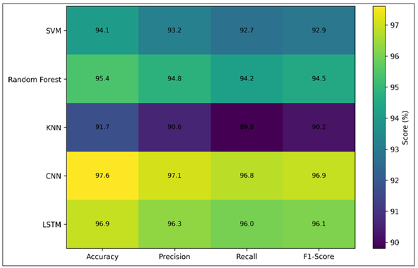
Figure 4 Heatmap Representation of Model Evaluation Metrics
The other factor of assessing machine learning models also relates to the trade off between the accuracy of the classification and the cost of computing them. Although deep learning models generally offer more accuracy in detection, the models have extended training durations since they are more complex in nature. Figure 5 represents the relationship between training time and the accuracy of classification wherein each point indicates a certain model in the study experiment. According to the scatter plot, CNN and LSTM models are the ones that have the highest level of accuracy, but they also consume more resources at the training stage. Random Forests and SVM models represent a positive tradeoff between the performance of training and the classification accuracy, and thus are appropriate in systems with limited computational resources.
Figure 5

Figure 5 Accuracy–Efficiency Trade off Among Classification Models
The ECG analysis systems are also evaluated based on the variety and quality of the datasets on which the model is trained and evaluated. The variability of the different datasets can be in terms of signal sampling frequency, number of recorded patients and duration of recorded signals. These properties determine the extent of generalization of trained models to clinical settings in the real world. The distribution of the dataset used in the study is shown in Figure 4 which is a bubble chart that explains the correlation between the sampling frequency and the quantity of the records in the popular ECG datasets. The bigger bubbles denote bigger datasets including more patient recordings, which mean that machine learning algorithms have more training data. The graphical representation demonstrates that big data like the PhysioNet challenge dataset offer a large amount of data on which to train powerful classification models.
Figure 6
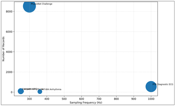
Figure 6 Dataset Landscape for ECG Signal Analysis
The general experimental findings can support the fact that the use of machine learning algorithms and the integration of visual analytics can greatly enhance the automated classification of the ECG.
9. Discussion
The outcomes of the carried out experimental evaluation suggest the opportunities of machine learning techniques to contribute to the effectiveness and precision of ECG signal examination. The potentials of the automated classification models were enormous in the detection of abnormal cardiac patterns of different ECG recordings. The results confirm that computational intelligence applications can be handy in the clinical diagnosis processes. The interpretability of the automated diagnostic systems can also be improved with the help of visual analytics and machine learning algorithms. ECG images presented graphically can allow clinicians to observe the trends of the waveforms concerning known abnormalities. This openness helps to gain the trust of the automated diagnosis tools, and also it allows the community of decision-making between the human experts and the computer systems. Irrespective of the good results, there are several issues in developing useful ECG diagnostic models. It is a research problem of interest to make sure that trained models are effective in various groups of patients. Hybrid machine learning models which entail the combination of deep learning models with the traditional signal processing models can be the basis of future studies. The technology of wearable healthcare has also evolved and as a result, real-time ECG monitoring systems can provide an opportunity to analyze cardiac signals even when there is no clinical environment. The future development of intelligent ECG diagnosis tools can transform the field of cardiac care by providing the opportunity to detect the abnormalities at the earliest stages and raise the accessibility of medical diagnosis.
10. Conclusion
Computer and automatized based analysis of electrocardiogram signals has become increasingly important in the improvement of quality and efficiency of cardiovascular diseases diagnosis. Having heart defects detected early helps the clinicians to undergo early medical treatment and reduce the probability of negative complications. The experiment in this paper developed a holistic model that combines the two visual analysis procedures and machine-based learning algorithms to detect the existence of cardiac abnormalities in ECGs. The proposed system will incorporate various algorithmic procedures, including signal preprocessing, feature extraction, visual cardiac pattern interpretation, and machine learning classification, which will enable its use in the area of the trustworthy detection of abnormal cardiac disorders. The experimental results showed that machine learning model combination can positively increase the accuracy of ECG signal classification by a significant margin. The traditional approaches to machine learning, such as Support Vector Machines and Random Forest classifiers, performed effectively when it comes to the identification of abnormal patterns in ECG. The specific type of deep architecture classification models, which were Convolutional Neural Networks and Long Short-Term Memory networks, increased the precision of classification due to the automatic learning of the representation of complex signals based on ECG waves. The results showed that deep learning models yielded the highest diagnostic results and the level of accuracy was more than ninety-seven percent. Visual analytics was also used to interpret the automated diagnostic framework. Graphical representations were represented by ECG waveforms, performance heatmaps, and the visualization of dataset distributions and helped to demonstrate the behavior of classification models and understand abnormal cardiac signal patterns in a more competent manner. The methods of visualizing data have a basis of clinical interpretation because they seek to provide intuitive insights of machine learning predictions. The created framework demonstrates the way in which the smart biomedical signal analysis systems can be utilized in the modern healthcare environment. Automated ECG diagnostic devices can be used to aid medical practitioners identify the existence of abnormalities in the heart more efficiently, in addition to reducing diagnostic workload and, therefore, enabling medical practitioners to observe their patients at all times. These systems would be even more practical when wearable health sensor and remote medical care services are added, which would not only enhance the possibility to realize the timely identification of any problem but would also make cardiac care services more accessible. More research in machine learning based ECG detectors would assist in development of more accurate, interpretable and scalable diagnostic tools in health care.
CONFLICT OF INTERESTS
None.
ACKNOWLEDGMENTS
None.
REFERENCES
Attia, Z. I., Harmon, D. M., Behr, E. R., and Friedman, P. A. (2021). Application of Artificial Intelligence to the Electrocardiogram. European Heart Journal, 42(47), 4717–4730. https://doi.org/10.1093/eurheartj/ehab649
Ayano, Y. M., Schwenker, F., Dufera, B. D., and Debelee, T. G. (2023). Interpretable Machine Learning Techniques in ECG-Based Heart Disease Classification: A Systematic Review. Diagnostics, 13(1), 111. https://doi.org/10.3390/diagnostics13010111
Basco, K. J., Singh, A., Nasef, D., Hartnett, C., Ruane, M., Tagliarino, J., Nizich, M., and Toma, M. (2025). Electrocardiogram Abnormality Detection Using Machine Learning on Summary Data and Biometric Features. Diagnostics, 15(7), 903. https://doi.org/10.3390/diagnostics15070903
Chong, L., Husain, G., Nasef, D., Vathappallil, P., Matalia, M., and Toma, M. (2025). Machine Learning Strategies for Improved Cardiovascular Disease Detection. Medical Research Archives, 13(1), 1–16. https://doi.org/10.18103/mra.v13i1.6245
Darmawahyuni, A., Nurmaini, S., Tutuko, B., Rachmatullah, M. N., Firdaus, F., Sapitri, A. I., Islami, A., Marcelino, J., Isdwanta, R., and Perwira, M. I. (2024). An Improved Electrocardiogram Arrhythmia Classification Performance with Feature Optimization. BMC Medical Informatics and Decision Making, 24, 412. https://doi.org/10.1186/s12911-024-02822-7
Denysyuk, H. V., Pinto, R. J., Silva, P. M., Duarte, R. P., Marinho, F. A., Pimenta, L., Gouveia, A. J., Gonçalves, N. J., Coelho, P. J., Zdravevski, E., et al. (2023). Algorithms for Automated Diagnosis of Cardiovascular Diseases Based on ECG Data: A Comprehensive Systematic Review. Heliyon, 9(7), e13601. https://doi.org/10.1016/j.heliyon.2023.e13601
Feyisa, D. W., Debelee, T. G., Ayano, Y. M., Kebede, S. R., and Assore, T. F. (2022). Lightweight Multireceptive Field CNN for 12-lead ECG Signal Classification. Computational Intelligence and Neuroscience, 2022, 8413294. https://doi.org/10.1155/2022/8413294
Goettling, M., Hammer, A., Malberg, H., and Schmidt, M. (2024). xECGArch: A Trustworthy Deep Learning Architecture for Interpretable ECG Analysis Considering Short-Term and Long-Term Features. Scientific Reports, 14, 13122. https://doi.org/10.1038/s41598-024-63656-x
He, Y., Zhou, Y., Qian, Y., Liu, J., Zhang, J., Liu, D., and Wu, Q. (2025). Cardioattentionnet: Advancing ECG Beat Characterization with a High-Accuracy and Portable Deep Learning Model. Frontiers in Cardiovascular Medicine, 11, 1473482. https://doi.org/10.3389/fcvm.2024.1473482
Jandera, A., Petryk, Y., Muzelak, M., and Skovranek, T. (2025). ECG Signal Analysis and Abnormality Detection Application. Algorithms, 18(11), 689. https://doi.org/10.3390/a18110689
Liu, X., Wang, H., Li, Z., and Qin, L. (2021). Deep Learning in ECG Diagnosis: A Review. Knowledge-Based Systems, 227, 107187. https://doi.org/10.1016/j.knosys.2021.107187
Pande, P. K., Khobragade, P., Ajani, S. N., and Uplanchiwar, V. P. (2024). Early Detection and Prediction of Heart Disease with Machine Learning Techniques. In Proceedings of the 2024 International Conference on Innovations and Challenges in Emerging Technologies (ICICET) (1–6). IEEE. https://doi.org/10.1109/ICICET59348.2024.10616294
Rahman, M. M., Rivolta, M. W., Badilini, F., and Sassi, R. (2023). A Systematic Survey of Data Augmentation of ECG Signals for AI Applications. Sensors, 23(11), 5237. https://doi.org/10.3390/s23115237
Śmigiel, S., Pałczyński, K., and Ledziński, D. (2021). ECG Signal Classification Using Deep Learning Techniques Based on the PTB-XL Dataset. Entropy, 23(9), 1121. https://doi.org/10.3390/e23091121
Varagani, S. H. N. S. (2026). Generative AI in Healthcare: Applications, Architecture, Implementation, and Benefits. International Journal of Advanced Computer Engineering and Communication Technology, 14(2), 53–63. https://doi.org/10.65521/ijacect.v14i2.1525
|
|
 This work is licensed under a: Creative Commons Attribution 4.0 International License
© ShodhKosh 2026. All Rights Reserved.